Posts

Abdominal Aortic Aneurysm: An Overview

Image
What is an Abdominal Aortic Aneurysm (AAA) ?       The aorta is the largest artery in the body and runs from the heart down to your chest and abdomen. Over many years, the walls  of the aorta may be weakened from the continuous pressure of blood flow, resulting in a bulge or swelling of the blood vessel known as an aortic aneurysm. The abdominal aortic aneurysm (also known as AAA, or ‘’triple A’’), is most commonly located just above your belly button       Due to the fact that these aneurysms result from the weakening of the aorta’s walls over time, this condition is typically found in the older age groups, in particular, older men aged over 65 years old. It is recommended that men of reaching this age or with a family history of AAA or a history of smoking receive a screening for this condition, as these aneurysms will gradually enlarge as time passes and this will often occur without patients experiencing symptoms. An AAA is commonly found unintentionally during a rout

All About Ultrasound

Image
What is an ultrasound and why do I need one?       An ultrasound is a painless, quick scan that is performed to allow your doctor to visualize the aorta. In this way, the ultrasound can confirm the presence of an aneurysm and also, very importantly, indicate the size of your aorta and of the aneurysm which is crucial.    The normal diameter for an aorta of a healthy adult male is 3cm. If a segment of the abdominal aorta widens to over 3cm it is considered to be an aneurysm. However, if this segment is under 5.5cm in width, the aneurysm most likely need not be operated on and should simply be monitored on a yearly basis if between 3-4.5cm and on a quarter yearly basis if between 4.5- 5.4 cm.      In this way, having an Ultrasound determines the course of action and treatment that   should be undertaken when you have an abdominal aortic aneurysm (AAA) and is thus very important.   How does ultrasound imaging work?    Ultrasound imaging involves sending hig

Ultrasound: How to Prepare and What to Expect

Image
 How should I prepare for my ultrasound?      There is little preparation required for an abdominal ultrasound in your test for an AAA. You may need to avoid eating for eight to 12 hours before the test and avoid drinking fizzy drinks and water during this time as this can cause gas in the intestinal tract which interferes with image quality. You should ask your doctor whether or not they require you to fast.   What should I expect on the day ?      When you are in the scanning room, you may be asked to change into a gown and to remove any accessories and jewellery. A gel will be placed over the area being scanned. This is to remove air pockets and to ensure that the transducer is in continuous contact with your skin. The gel is usually cold. Don’t worry; the gel is water-based and is easy to remove from both your skin and your clothing. The ultrasound technician will move the transducer across your skin. It is important that you lie still in order to prevent the

Ultrasound: Intrepreting Your Results

Understanding your Results What do my results mean ?       I f the results of your ultrasound show that you do in fact have an AAA, the next step will be to determine the size of the aneurysm to understand its severity. Possible Results: All Segments of the Abdominal Aorta less than 3cm in diameter      This is a perfectly normal result and indicates that you do not, in fact, have an abdominal aortic aneurysm (AAA).  As it takes a very long time to grow, there is little chance that you will develop one after the age of 65 and therefore no follow-up screenings or scans will be necessary. A Segment of the Abdominal Aorta between 3-5.5cm in diameter       If your aneurysm is found to be within these measurements, you have a small or medium aneurysm. This means it’s in or around the same size as an earphone. Aneurysms of this size are not of high risk and because of this, no treatment is required just yet. You will, however, have to attend various follow

Other Scans for AAA: CT

Image
What is a CT scan and why do I need one?      A computerized tomography (CT) scan combines a series of X-ray images captured from different angles and utilizes computer processing to produce image slices. It gives clearer images and more depth to the bones, vessels, and organs.therefore, it is used to scan patients that show symptoms and are suspected to have AAA.   Contrast-Enhanced CT (CECT) may also be used to examine patients that do not show symptoms but have an aneurysm bigger than 5.5cm. How does CT scanning work?      A typical abdominal CT scan takes about 10 to 30 minutes. you will be asked to lie down on the procedure table. With the use of a remote control from a separate room, the technician will move the table into the CT machine, which looks like a giant doughnut comprised of metal and plastic. You will most likely go through the machine several times before the exam is complete. The X-ray tubes in the machine will emit radiation beams that will go th

Other Scans for AAA: MRI

Image
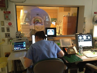
What is an MRI and why do I need one?     MRI (magnetic resonance imaging) is another imaging procedure that you might encounter with an AAA. It uses magnetic fields to obtain a clear picture of internal organs. It is a particularly effective exam as it allows the aorta to be imaged without the use of contrast agents or ionising radiation. It is used as it often offers better imaging of the aorta branch vessels than other exams and may also be used when it is unadvisable for the patient to receive ionised contrast used in CT.   MRI control room (Source:  https://basicsofpediatricanesthesia.com/30-mri-control-room/  ) How does MRI imaging work?      An MRI uses a magnetic field and a radio-frequency current to stimulate the protons in your body, causing them to strain against the pull of the magnetic field. MRI sensors create images by detecting the energy released as the protons in your body realign with the magnetic field after the radio-freque